
#82 Intro to Neurocritical Care w/ Dr Casey Albin
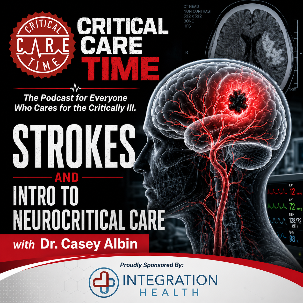
We are proud to bring you guys our first of several episodes on Neurocritical Care! On this episode - our introductory show - we had the pleasure of hosting Dr. Casey Albin a neurologist & neurointensivist who introduces the topic of neurocritical care with a special focus on basic tenets of stroke management. What type of imaging should you order? What do you do with anticoagulation? Who get's seizure prophylaxis? We cover all this and more with Dr. Albin. Please take a listen and let us know what you think and what else you want to hear from us!
Our Guest
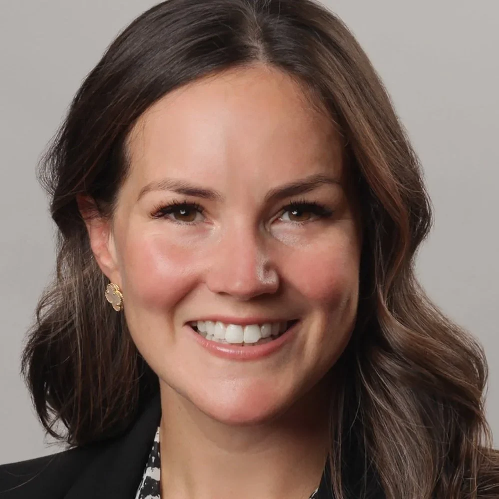
Casey Albin, MD
Associate professor of Neurology & Neurosurgery in the Division of Neurocritical Care at Emory University
Case 1: Elevated Intracranial Pressure
The case: A 28-year-old after a motor vehicle collision arrives intubated, GCS 6T, with a sluggish right pupil. CT shows a traumatic subdural hematoma, contusions, effaced cisterns, and a 7 mm midline shift.
The Monro-Kelli Doctrine: The skull is a fixed-volume container composed primarily of brain tissue, blood, and cerebrospinal fluid. Early compensation occurs by shifting CSF and venous blood out of the cranial vault, but once reserve is exhausted, small increases in volume produce dramatic increases in ICP (intracranial pressure).
Initial bedside management includes:
Elevate the head of the bed
Maintain the neck in neutral position
Avoid jugular venous compression (e.g. C-collar)
Provide adequate analgesia and sedation
Correct hypoxia and hypercapnia
Treat fever, seizures, and hyponatremia
Intubate when necessary to control oxygenation and ventilation
Interventions like Hypertonic saline and mannitol reduce cerebral edema and lower ICP, although they have not consistently demonstrated improved long-term neurologic outcomes. Brief hyperventilation remains a temporizing maneuver rather than definitive therapy.
ICP, MAP and CPP - Remember the fundamental equation:
CPP (Cerebral perfusion pressure) = MAP (mean arterial pressure) − ICP
An elevated ICP threatens both immediate herniation and cerebral perfusion. Most severe traumatic brain injury protocols target:
ICP <22 mmHg
CPP approximately 60–70 mmHg (individualized)
MAP often >75–80 mmHg to maintain adequate CPP
Cushing's Triad — and a Common Mistake
Hypertension, bradycardia, and agonal respirations. The physiology: elevated ICP limits diastolic cerebral flow, so the brain becomes dependent on systole for perfusion — and more time is spent in systole with a faster heart rate.
Do not reflexively "normalize" that reflex tachycardia with a beta blocker; you'd be removing the compensatory mechanism keeping the brain perfused.
Myth-Busting: Does an IJ Central Line Raise ICP?
Observational data out of Emory found no significant change in ICP before vs. after IJ central line placement. Myth: busted.
External Ventricular Drains
An EVD provides two important functions:
Continuous ICP monitoring
Therapeutic CSF drainage
Compared with intraparenchymal bolts, EVDs also allow treatment of hydrocephalus and are particularly valuable in subarachnoid hemorrhage, intraventricular hemorrhage, meningitis, and severe traumatic brain injury.
Case 2: Acute Ischemic Stroke
The case: A 72-year-old with atrial fibrillation develops aphasia and right hemiplegia 70 minutes before arrival, with a high NIHSS. CT shows no hemorrhage; CTA reveals a left M1 occlusion.
Management begins with a simple question: Is there blood?
A noncontrast CT excludes hemorrhage, after which CTA evaluates for large vessel occlusion and CT perfusion can identify salvageable penumbra beyond traditional time windows.
For patients with LVO:
Administer thrombolysis when appropriate.
Proceed rapidly to mechanical thrombectomy.
At non-thrombectomy centers, "drip and ship" remains the standard approach.
Last Seen Well vs. the Tissue-Based Clock
The classic 4.5-hour thrombolysis window has been pushed by newer trials using perfusion imaging (CT perfusion, CTP) to identify salvageable penumbra vs. already-infarcted core — shifting decision-making from a pure time clock to a tissue clock in select patients.
Thrombolysis & Thrombectomy Pathways
Thrombectomy-capable center: give TNK and go straight to thrombectomy in parallel
Non-thrombectomy-capable center: "drip and ship" — give TNK, then transfer for thrombectomy
ASPECTS score (noncontrast CT, 0 = diffuse early ischemic change, 10 = normal): a score >3 generally favors proceeding to thrombectomy (salvageable tissue still present)
Thrombectomy eligibility: essentially all M1 occlusions; basilar occlusions when NIHSS >10; M2 occlusions more selectively — recent trials didn't show a clear functional benefit in M2s, though patient selection in those trials was imperfect
After Reperfusion — What Does the ICU Team Do?
Rule out hemorrhagic reperfusion injury
Extubate if the patient was left intubated — most post-thrombectomy patients actually do great
Watch for reocclusion and access-site complications
Avoid post-procedure hypertension (target SBP <160)
Start the etiology workup
Optimize the recovery environment: goldilocks temperature and glucose control
Secondary Prevention
Above-the-neck disease (small vessel disease, intracranial atherosclerosis) → antiplatelet therapy (DAPT)
Cardiac source (atrial fibrillation, paradoxical embolus) → anticoagulation
Learning Points
The first decision is always ischemic vs. hemorrhagic; treatments diverge immediately from there.
Noncontrast CT rapidly excludes hemorrhage; vascular imaging identifies the LVO.
Acute ischemic stroke systems prioritize rapid reperfusion, thrombolytic and thrombectomy eligibility, BP/glucose/temperature control, dysphagia screening, and surveillance for edema or hemorrhagic transformation.
LVO patients frequently need ICU-level care for airway management, blood pressure strategy, reperfusion complications, malignant edema, and post-procedure neuro monitoring — but many, even with large strokes, tolerate extubation better than you'd expect.
Case 3: Intracerebral Hemorrhage
The case: A 64-year-old on apixaban presents with acute left-sided weakness, vomiting, and somnolence. BP is 218/112. CT shows a right basal ganglia ICH with intraventricular extension and early hydrocephalus.
A Field That's Moving Past Nihilism
ICH has historically been treated with therapeutic nihilism, but the evidence base is shifting — through blood pressure control, anticoagulation reversal, and a re-examination of surgical candidacy (more on ENRICH below).
First ICU Priorities
Airway control comes first, followed immediately by balancing blood pressure reduction (to limit hematoma expansion) against maintaining adequate cerebral perfusion pressure.
Blood Pressure Targets
Initial SBP <220: target ~140, with a safe/reasonable range of 130–150. Pushing below 130 may cause harm.
Initial SBP ≥220: target <180 acutely, then <160 the following day.
Anticoagulation in the DOAC Era
Most hypertensive ICH patients on a DOAC for a good indication still ultimately benefit from resuming anticoagulation once stabilized — this is a risk-benefit conversation, not a blanket stop. Extra caution is warranted in elderly patients where cerebral amyloid angiopathy (CAA) is the likely underlying cause, given the higher recurrence risk.
Reversal: for DOAC dosing within the last 48 hours, reverse with 4-factor PCC (e.g., Kcentra, 25–50 IU/kg).
When Does Neurosurgery Become Urgent?
Roughly 30–80 cc of cortical/basal ganglia blood is the range where the ENRICH trial demonstrated a functional recovery benefit from minimally invasive evacuation — a genuine shift after decades of negative surgical trials in ICH.
Surgical Options at a Glance
EVD for obstructive hydrocephalus
Suboccipital craniectomy for cerebellar bleeds (best evidence of the surgical options)
Decompressive craniectomy as a life-saving measure; less robust data for other lesion types
Cortical bleeds may derive some surgical benefit per ENRICH
Seizure Prophylaxis
Spontaneous ICH alone is unlikely to cause seizures; cortical bleeds carry higher seizure risk. Reach for EEG when there's altered mental status or fluctuating exam, rather than reflexive prophylactic antiseizure medication. (Traumatic ICH is the exception — 7 days of seizure prophylaxis is standard there.)
Learning Points
ICH priorities: confirm the diagnosis, stop expansion, reverse anticoagulation, control blood pressure smoothly, identify hydrocephalus/IVH, and monitor closely for deterioration.
For mild-to-moderate ICH with SBP 150–220, target ~140 mmHg; maintaining 130–150 is considered safe, but pushing below 130 may be harmful.
Anticoagulant-associated ICH requires immediate discontinuation and rapid reversal.
IVH with hydrocephalus and depressed consciousness is an EVD/neurosurgical problem.
Don't use prophylactic antiseizure medications without evidence of seizures — reserve EEG for fluctuating mental status or suspected seizure activity.
Case 4: Subarachnoid Hemorrhage
The case: A 51-year-old reports the "worst headache of life," collapses, then improves. CT shows diffuse subarachnoid blood; CTA identifies an anterior communicating artery aneurysm.
Aneurysmal SAH is far more than simply "blood on the CT." It's a multisystem neurocritical illness — an arterial bleed occurring outside the brain parenchyma itself, most commonly from an anterior or posterior communicating artery aneurysm.
What Kills Early
Rebleeding, acute hydrocephalus, elevated ICP, and cardiopulmonary complications — which is why early priorities are airway support as needed, blood pressure control before the aneurysm is secured, recognition of hydrocephalus (with EVD as needed), and prompt aneurysm repair. Endovascular coiling is increasingly taking over from surgical clipping.
Nimodipine — Not a Class Effect
Nimodipine specifically (not calcium channel blockers as a class) improves functional recovery in SAH. Target euvolemia — hypervolemia is not preventive and shouldn't be pursued prophylactically.
The Late Course: Delayed Cerebral Ischemia (DCI)
This is where SAH diverges sharply from the early-mortality story. DCI is a clinical finding; vasospasm is a biomarker — they're related but not synonymous, and it's a distinction worth internalizing.
Daily transcranial Dopplers (TCDs) to monitor for vasospasm
If vasospasm develops, treat with nicardipine or milrinone
Once the aneurysm is secured, permissive hypertension (letting the patient auto-regulate upward) is reasonable
Magnesium may help via vasorelaxation — goal Mg >2
Q1h neuro checks for roughly the first 48 hours
Learning Points
SAH is a multisystem neurocritical illness, not just "blood on CT."
ACoM and PCoM aneurysms are the most common sources.
Early priorities: airway as needed, BP control before aneurysm securement, hydrocephalus recognition with EVD as needed, aneurysm repair, nimodipine, euvolemia, and DCI surveillance.
Coiling is increasingly favored over clipping as endovascular therapy advances.
Deterioration days later is a classic teaching scenario — think vasospasm/DCI, hydrocephalus, seizures, hyponatremia, fever, and cardiac injury.
Closing Thoughts
Casey's book, Acute Neurology Survival Guide, is a great bedside companion for exactly these scenarios. Find her on Instagram at @casey_albin.
Sponsored by Integration Health

-
-
The Physiologically Difficult Airway
The Anatomically Difficult Airway Part 1
The Anatomically Difficult Airway Part 2
-