
#84 Acid-Base w/ Dr Sara Crager
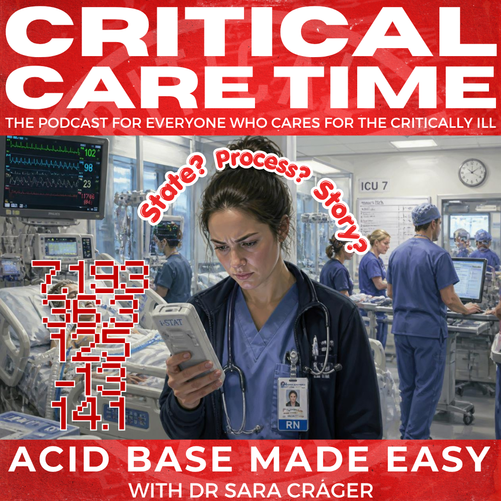
Returning guest Dr. Sara Crager joins us again on Critical Care Time, this time to tackle one of the most important, and most commonly overcomplicated, topics in critical care: acid–base physiology. Rather than treating acid–base as a collection of equations and compensation formulas, Sara walks us through her practical State → Process → Story framework for rapidly interpreting blood gases and electrolytes at the bedside. We discuss how to distinguish acidemia from acidosis, interpret CO₂ in the context of bicarbonate, use the anion gap and strong ion difference to uncover mixed metabolic disorders, recognize the importance of chloride and albumin, and understand why a “normal” pH—or a “normal” CO₂—may be anything but normal. Along the way, we work through several real-world ICU cases involving chronic hypercapnia, DKA, vomiting, septic shock, renal failure, saline-induced hyperchloremia, and hypoalbuminemia. The goal isn’t more acid–base math. It’s a better mental model and a faster way to understand what your patient’s numbers are actually telling you.
Our Guest

Dr. Sara Crager, MD
Emergency Physician and Intensivist. Faculty at UCLA in the Departments of Emergency Medicine and Anesthesia Critical Care
Acid–Base 2.0: State, Process, Story
Acid–base physiology is often taught as a collection of equations to memorize. In this episode, Dr. Sara Krieger returns to Critical Care Time with a more clinically useful approach: think of acid–base abnormalities as multiple simultaneous processes, then rapidly determine which ones are operating in your patient.
Sara's bedside framework is simple:
State → Process → Story
1. STATE: What is the pH?
Start with the patient's current state:
Acidemia: pH < 7.35
Alkalemia: pH > 7.45
Normal pH: does not mean normal acid–base physiology.
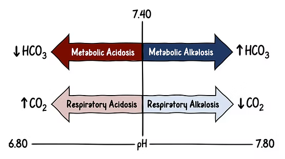
An -emia describes the patient's current state; an -osis describes a process producing that state. Multiple opposing processes can therefore produce a completely normal pH.
Think of acid–base physiology as a vector sum of four possible processes:
Metabolic acidosis ↔ Metabolic alkalosis
Respiratory acidosis ↔ Respiratory alkalosis
The measured pH reflects their net effect.
2. PROCESS: What processes are occurring?
Work through respiratory and metabolic processes separately.
Respiratory: Interpret CO₂ in the context of HCO₃⁻
Never decide that a PaCO₂ is "normal" simply because it falls within the laboratory reference range. A PaCO₂ of 40 may represent severe respiratory acidosis in a patient whose HCO₃⁻ is 10.
Acute metabolic acidosis: the quick compensation check
For a metabolic acidosis:
Expected PaCO₂ ≈ the last two digits of the pH
Example:
pH 7.24
PaCO₂ 24 mmHg
HCO₃⁻ 10 mEq/L
→ appropriate respiratory compensation.
If PaCO₂ is higher than expected → additional respiratory acidosis.
If PaCO₂ is lower than expected → additional respiratory alkalosis.
This shortcut becomes unreliable with very severe acidemia (roughly pH <7.10), where respiratory compensation reaches physiologic limits.
Chronic respiratory acidosis: the 10/4 rule
For every chronic 10-mmHg rise in PaCO₂, expect approximately a 4-mEq/L rise in HCO₃⁻.
This lets you estimate a chronically hypercapnic patient's baseline PaCO₂ rather than assuming every elevated CO₂ represents acute respiratory failure.
The goal isn't necessarily to make their PaCO₂ "normal" — it's to determine what is normal for that patient.
And remember: compensation never overcompensates. If the numbers go beyond expected compensation, there is another primary acid–base process.
Metabolic: Stop thinking of HCO₃⁻ as a single process
HCO₃⁻ is better understood as the net result of several metabolic processes:
Anion-gap metabolic acidosis ↓
Non-anion-gap metabolic acidosis ↓
Metabolic alkalosis ↑
These processes can coexist and partially cancel one another.
Sara therefore interprets HCO₃⁻ using two numbers:
Anion Gap
AG = Na − Cl − HCO₃
A high anion gap means an anion-gap metabolic acidosis is present, regardless of the pH or HCO₃⁻.
Where there's a gap, there's an acidosis.
Strong Ion Difference
For bedside purposes:
SID ≈ Na − Cl
Normal ≈ 38–40 mEq/L
Low SID (<38–40) → non-anion-gap metabolic acidosis
High SID (>40) → metabolic alkalosis
Importantly, this is about chloride relative to sodium, not the absolute chloride concentration. A chloride of 115 may be perfectly appropriate if the sodium is proportionately elevated.
This provides a simple alternative to wrestling with delta-delta calculations: calculate Na − Cl, then subtract HCO₃⁻ to get the anion gap. Those two quick calculations reveal much of the patient's metabolic acid–base physiology.
Why chloride matters
The body prioritizes electroneutrality. Sodium and chloride are the major strong ions in plasma, while bicarbonate can change to preserve charge balance.
Thus:
↑ chloride relative to sodium → ↓ SID → ↓ HCO₃⁻ → metabolic acidosis
↓ chloride relative to sodium → ↑ SID → ↑ HCO₃⁻ → metabolic alkalosis
This is why saline-induced hyperchloremia produces a non-gap metabolic acidosis, while chloride depletion from vomiting or diuretics contributes to metabolic alkalosis.
Don't forget albumin
Albumin accounts for much of the normal anion gap. In severe hypoalbuminemia, a seemingly "normal" AG may therefore conceal an anion-gap acidosis.
A useful correction:
Corrected AG ≈ measured AG + 2.5 × (4 − albumin)
Or as a quick bedside approximation discussed in the episode:
Expected normal AG ≈ 3 × albumin
This becomes especially important in critically ill patients with cirrhosis, malnutrition, or profound hypoalbuminemia.
3. STORY: Make the numbers explain the patient
Finally ask:
What clinical story explains all of these processes simultaneously?
This is where acid–base interpretation becomes clinically useful rather than an exercise in arithmetic.
A patient with DKA, for example, may have:
AG metabolic acidosis → ketoacidosis
Metabolic alkalosis → vomiting/chloride depletion
Respiratory alkalosis → pain, sepsis, pregnancy, etc.
The resulting pH may even be normal. In the episode's DKA case, pH 7.39 concealed three simultaneous acid–base disorders.
Similarly, the same ABG can mean very different things depending on the electrolytes and clinical context. Don't interpret a blood gas in isolation from the chemistry panel.
Take-Home Points
State → Process → Story.
-Emia is a state; -osis is a process. A normal pH does not exclude major acid–base abnormalities.
Interpret PaCO₂ in the context of HCO₃⁻, not against the laboratory reference range.
In metabolic acidosis, expected PaCO₂ ≈ the last two digits of the pH.
For chronic respiratory acidosis, remember 10/4: HCO₃⁻ rises ~4 for every chronic 10-mmHg rise in PaCO₂.
Think of HCO₃⁻ as the net vector sum of metabolic processes.
AG = Na − Cl − HCO₃⁻ identifies anion-gap acidosis.
SID ≈ Na − Cl; normal is roughly 38–40. Low SID indicates non-gap acidosis; high SID indicates metabolic alkalosis.
Correct the anion gap for severe hypoalbuminemia.
Don't make the numbers pretty — figure out the patient's story.
-
-
Metabolic Acidosis
-
Sara's acid–base resources, graphics, and pocket cards: