
#83 B2B Pulse Oximetry
[blurb]
About the B2B series
The Back to Basics (B2B) series takes a piece of equipment we use — and take for granted — every day in the ICU and breaks it down at three levels:
101 — foundational knowledge for people new to the ICU (medical students, nursing students)
201 — the "why behind the what" for people who already know the device
301 — advanced tricks, edge cases, and myth-busting for experienced clinicians
This episode: pulse oximetry, the free, continuous, non-invasive vital sign we all rely on and almost never think critically about.
101 — The basics
When to use pulse oximetry?
Essentially always in the ICU — it's free, continuous, non-invasive data. But two caveats to remember before you slap on a probe and walk away:
Alarms aren't free. Too many nuisance alarms causes alarm fatigue, which means real alarms get ignored — and it disrupts patient sleep, which contributes to delirium.
Measurement drives action, and not all action helps. A low SpO₂ reading in the hospital usually triggers more supplemental oxygen. That's exactly why picking the right target matters.
Choosing the right SpO₂ target
More is not better. Medicine selects for perfectionists — people who want everyone's sat at 100%. That instinct is wrong here. Patients do not need, nor do they benefit from, 100% SpO2. Use reasonable evidence-based targets instead:
Reasonable default targets:
| Population | Target SpO₂ | |
|---|---|---|
| Most patients | 92–96% | |
| COPD / chronic hypercarbia | 88–92% | |
| Pregnancy | >95% (physiologic rationale — more than outcome-trial-based) |
(Don’t worry we'll debunk the "too much oxygen makes you stop breathing" myth in the 301 section.)
Why hyperoxia is a problem, not just neutral: In ARDS, acute MI/ACS, and stroke, hyperoxia has real signal for harm. And a "perfect" SpO₂ of 100% is deceptive — it could reflect a PaO₂ anywhere from 100 to 400+ mmHg. SpO₂ lacks resolution in cases where it matters the most.
The evidence trail on oxygen targets:
The AVOID trial (Circulation, 2015) randomized 441 non-hypoxic STEMI patients to supplemental oxygen vs. none and found high-dose supplemental oxygen was potentially harmful in normoxic STEMI patients, with increased myocardial injury and more recurrent MI. That's why there's no more "O" in MONA.
The Oxygen-ICU trial (JAMA, 2016, n=434) found a conservative oxygen strategy (SpO₂ 94–98%) was associated with markedly lower ICU mortality than a liberal one (SpO₂ 97–100%) — 11.6% vs. 20.2%, ARR 8.6%, NNT 11 — a striking effect size.
That mortality benefit didn't replicate in the larger ICU-ROX trial (NEJM, 2020, n≈1,000): conservative oxygen therapy (lower SpO₂ targets) did not significantly affect ventilator-free days compared with usual oxygen therapy — though a signal for harm from hyperoxia persisted in the broader literature.
The much larger UK-ROX trial (JAMA, 2025, n=16,500) found no mortality benefit from targeting a lower SpO₂ (~90%) versus usual care.
Bottom line: more oxygen definitely isn't better. We target normoxia, not maximal saturation.
Don't make big decisions with bad data
“Don’t draw vast conclusions from half vast data!”
Before you trust a number, check the waveform.
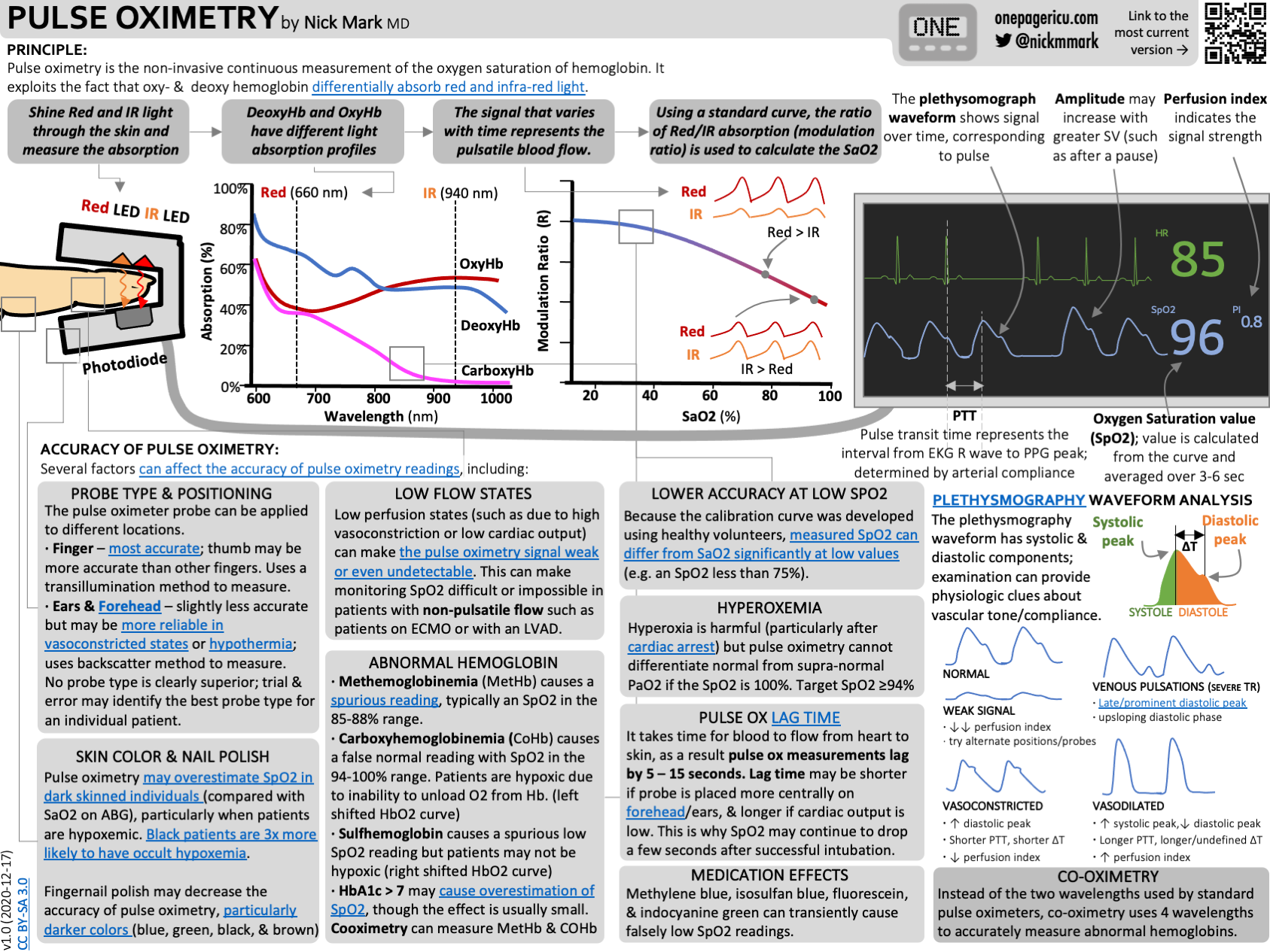
The plethysmograph ("pleth") is the visual pulse-ox waveform — it should look like a pulse, with clear peaks per beat. A bad waveform means bad data, full stop.
Some monitors display a perfusion index (PI) — the ratio of pulsatile to non-pulsatile blood flow, typically 0.02–20%. A PI of 4–6% suggests a strong signal; below ~1.0% (or 0.4%) suggests poor peripheral perfusion, cold extremities, or shock.
Cross-check your numbers. Compare the "blue HR" (derived from the pulse ox) against the "green HR" (from the EKG) — mismatches can reveal double-counting or under-detection on either monitor.
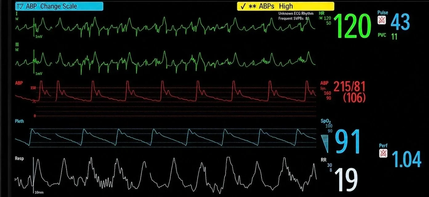
In this case the ECG HR (green) is 120 whereas the Pulse Ox HR (blue) is 43. Which is telling the truth? In this case the pulse ox is telling the truth as confirmed by comparing the arterial line and pleth tracings.
Where you can measure — and why signal quality varies
Pulse oximetry is perfusion-dependent: you need a good sensor in a good position over pulsatile capillary flow. Common sites: fingers/toes, ears, nose, forehead. In cold, vasoconstricted patients, a good signal anywhere can be hard to find.
The pulse ox is a time machine
You're seeing blood oxygenation as it was when that drop of blood left the heart — typically a 15–30 second lag, and it can stretch past a minute in heart failure or any state with a long circulation time. Lag also depends on probe site: less lag at the ear or forehead, more at the fingertip or toe.
201 — Beyond the basics
How pulse oximetry actually works
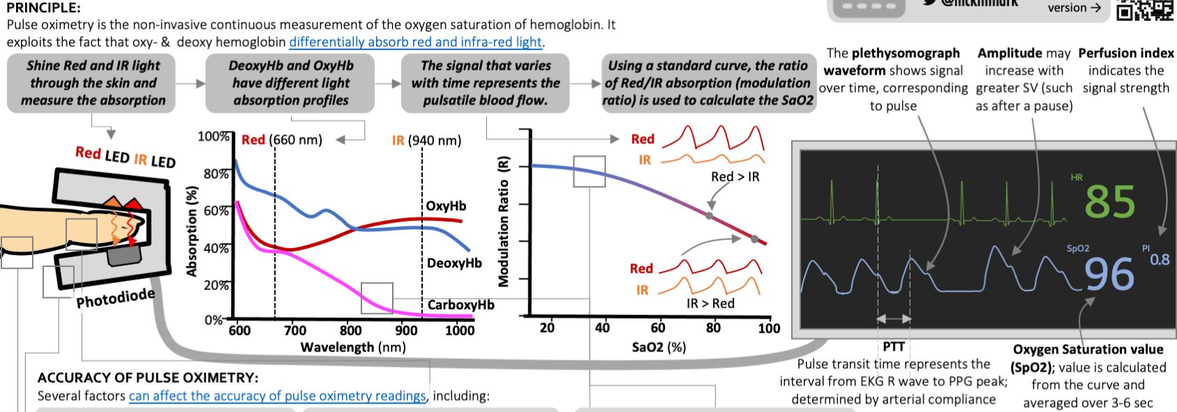
Pulse oximetry exploits a simple optical trick: oxygenated hemoglobin absorbs more infrared light (~940 nm) and lets more red light (~660 nm) through, while deoxygenated hemoglobin does the opposite. The device computes the ratio (R) of pulsatile absorption at those two wavelengths and maps it to an SpO₂ value using an empirical calibration curve built from human volunteer studies.
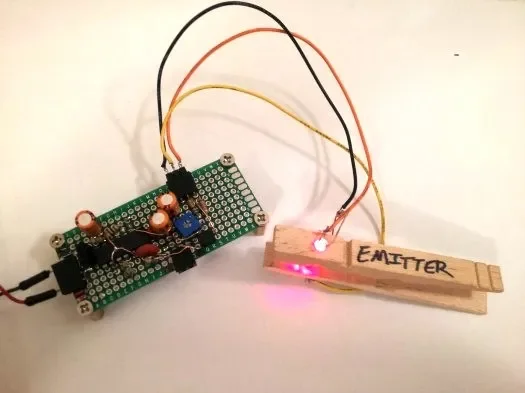
Fun side project: you can build a basic pulse oximeter yourself with off-the-shelf parts. Great kid science project or rainy weekend project. For example check this out.
A little history and a big problem
Pulse oximetry was originally built to detect hypoxemia in pilots and became a standard ICU monitoring tool in the 1980s. Today you can buy one on Amazon for under $20.
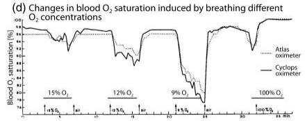
Effect of breathing different FiO2 concentrations on health volunteer SpO2 measurements. Normobaric hypoxemia was used to develop the non-invasive pulse oximetry measurement technique. Modified from Queresima et al, 2024
The original calibration studies used healthy young Caucasian male volunteers. Researchers placed arterial lines, had subjects breathe low-O₂/high-nitrogen gas mixtures to induce controlled hypoxia, and measured SaO₂ and SpO₂ simultaneously to build the calibration model — but ethics boards wouldn't allow dropping saturations far enough to calibrate below roughly 75%, so accuracy below that point is inherently less precise for everyone.
More importantly: because the original cohort was all Caucasian, the calibration introduced a racial bias — most pulse oximeters overestimate SpO₂ in darker-skinned individuals, by up to about 3%. This is bias in both senses of the word: statistically (a systematic measurement error) and socially (it produces unequal care). Because decisions about intubation, home oxygen, ECMO candidacy, and lung transplant listing are often driven by SpO₂ readings, a systematic error in measurement becomes a systematic disparity in treatment. It's also a broader argument for requiring diverse, representative populations in FDA device-testing standards.
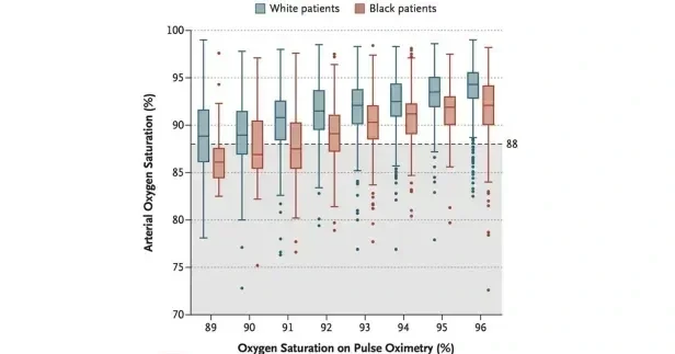
Systematic bias in pulse oximeter measurements tends to overestimate SpO2 in darker skinned individuals. From Sjoding et al, NEJM
Abnormal hemoglobins and other artifacts
| Hemoglobin variant | Blood color | Reads as | Common causes |
|---|---|---|---|
| Carboxyhemoglobin | Bright/cherry red | Falsely ~100% | Fires, engine exhaust, space heaters, grills, methylene chloride exposure |
| Methemoglobin | Chocolate brown | ~80% | Benzocaine, dapsone, bactrim, chloroquine, primaquine, nitrates, poppers, rare massive Tylenol overdose |
| Sulfhemoglobin | Greenish | Low 70s–80s (may be less dangerous — right-shifts the oxyhemoglobin curve) | Sulfasalazine, phenazopyridine, sumatriptan, industrial exposure (e.g., TNT workers) |
Other things that interfere with the pulse ox reading:
Nail polish — can throw off the signal.
Continuous-flow devices (ECMO, LVAD) — pulse oximetry needs pulsatile flow, so continuous-flow circulatory support may not register a signal at all.
Probe placement matters — right hand is important for peripherally cannulated VA-ECMO, and for pre-ductal measurement in pediatrics.
Prehospital/transport tip — covering the probe with a towel can help preserve signal quality, and be ready to explain a strange reading if you push methylene blue near a colleague unfamiliar with the effect.
Tissue oximetry (a related but different technique)
Pulse oximetry measures continuous arterial blood oxygenation (SpO₂). Tissue oximetry, typically using near-infrared spectroscopy (NIRS), instead measures regional oxygen saturation (rSO₂) in a mixed pool of arterial, capillary, and venous blood within a specific tissue bed — commonly brain or limb. It's especially useful for monitoring cerebral and limb perfusion during ECMO.
301 — Advanced: mechanisms, edge cases, and myths
Why is too much oxygen actually bad?
Beyond COPD, hyperoxia has several distinct downsides:
Causes coronary and cerebral vasoconstriction, potentially reducing blood flow despite a higher PaO₂.
Increases reactive oxygen species, driving mitochondrial dysfunction, lipid peroxidation, and inflammatory injury.
Produces absorption atelectasis: high FiO₂ washes nitrogen out of alveoli, so poorly ventilated units collapse as their oxygen gets absorbed.
Causes direct pulmonary toxicity at high FiO₂ with prolonged exposure.
Worsens reperfusion injury after cardiac arrest, stroke, or myocardial ischemia.
Conceals deterioration — a patient on excessive FiO₂ can sit at 100% SpO₂ while their underlying gas exchange is worsening.
Dries mucosa and adds unnecessary device-related discomfort.
And in COPD specifically: the classic "hypoxic respiratory drive" explanation is largely a myth. The bigger contributors to CO₂ retention with high-flow oxygen are the Haldane effect and worsening V/Q mismatch (loss of hypoxic pulmonary vasoconstriction redirects blood to poorly ventilated units).
Detecting abnormal hemoglobins: co-oximetry
Standard two-wavelength (red/IR) pulse oximetry can't distinguish these variants. Co-oximetry uses four (or more) specific wavelengths — commonly cited examples include 535, 585, 594, and 626 nm, though exact combinations vary by manufacturer — to differentiate oxyhemoglobin, deoxyhemoglobin, carboxyhemoglobin, and methemoglobin.
Methemoglobinemia
Mechanism: the heme iron is oxidized from ferrous (Fe²⁺) to ferric (Fe³⁺), which can't bind oxygen — causing both a functional anemia and a left shift of the oxyhemoglobin curve that impairs tissue oxygen unloading. Even one oxidized subunit in a hemoglobin tetramer impairs the whole molecule's unloading.
Causes: dapsone, topical anesthetics (benzocaine, lidocaine), antimalarials, industrial chemicals (nitrates/nitrites, aniline, dyes).
Treatment: IV methylene blue (1–2 mg/kg) reverses the oxidation; for G6PD deficiency or severe contraindications, vitamin C (ascorbic acid) is used instead.
Carboxyhemoglobinemia
Mechanism: CO binds the same site as O₂ with roughly 200–250x the affinity, so even trace CO exposure rapidly occupies hemoglobin. Like methemoglobin, it also left-shifts the curve, worsening oxygen unloading.
Causes: tobacco smoke, incomplete combustion (faulty furnaces, space heaters), car exhaust, methylene chloride.
Treatment: oxygen — possibly hyperbaric oxygen in severe cases.
Sulfhemoglobinemia
Mechanism: right-shifts the oxyhemoglobin curve (the opposite of the other two), which actually enhances peripheral oxygen release — making it physiologically less dangerous despite the alarming color.
Causes: sulfonamides, metoclopramide, phenacetin (banned in the early 1980s), aniline dyes.
Treatment: stop the offending drug. Because the binding is irreversible, transfusion is the only way to clear it.
[IMAGE: oxyhemoglobin dissociation curve showing left shift (met-, carboxyhemoglobin) vs. right shift (sulfhemoglobin)]
Cyanosis: useful but unreliable
Cyanosis requires 5 g/dL of deoxygenated hemoglobin to become visible. Because it's an absolute quantity, not a percentage, the saturation at which cyanosis appears depends heavily on total hemoglobin:
Polycythemic patient (Hgb 20) → visibly cyanotic even at ~75% saturation
Normal hemoglobin (Hgb 10) → cyanotic around 50% saturation
Anemic patient (Hgb 7) → may not look cyanotic until ~30% saturation
Cyanosis is a real sign of hypoxemia, but it's a late one and is hemoglobin concentration dependent — don't rely on it alone!
What the waveform tells you beyond "signal present"
The plethysmograph carries subtle physiologic information beyond signal quality:
Venous pulsations on the waveform can be a clue to tricuspid regurgitation.
Extreme vasoconstriction or vasodilation is visible in waveform shape.
Sometimes you can watch a patient's physiology change with resuscitation in real time just by watching the pleth.
Pulse Oximetry in LVAD Patients
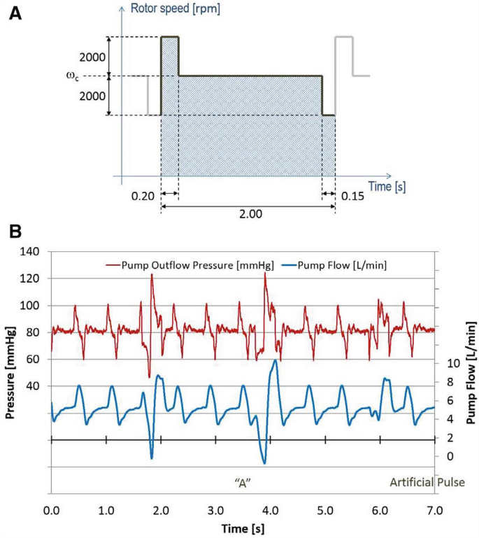
By varying the rotor speed every 2 seconds the HeartMate3 LVAD generates a pulsatile signal, allowing the pulse oximeter to work. Source: Peev & Salerno
LVAD quirk: the HeartMate 3 includes a built-in "artificial pulse" (intrinsic pulsatility) — the pump cycles about 30 times/minute, dropping RPM by 2,000 for 0.15 seconds then increasing by 4,000 for 0.20 seconds before returning to baseline. It exists to minimize blood stasis, improve pump washout, and prevent thrombus formation — but as a side effect, it can generate enough pulsatility to produce a pulse ox reading. Note the rate on the monitor won't necessarily be accurate.

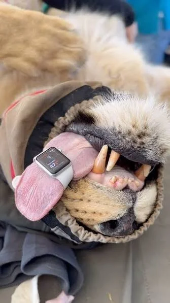
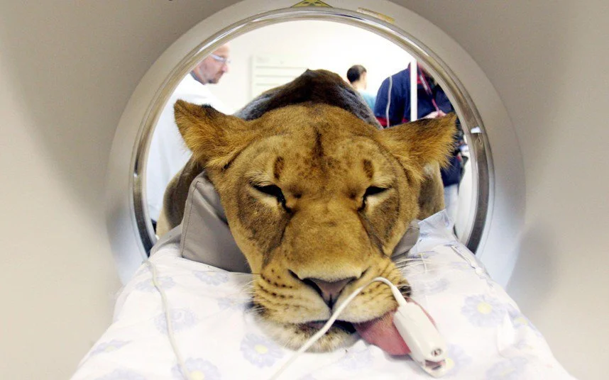
Does pulse ox work on non-humans?
Pulse oximetry works reliably in cats, dogs, and horses — but not universally:
White rhinoceros: under opioid immobilization, rhinos regularly show pulse-ox readings indicating severe hypoxemia — yet recover uneventfully, with normal arterial blood gases. Their hemoglobin simply doesn't read correctly on standard devices.
Birds (nucleated red blood cells) and reptiles (different hemoglobin structure) give less reliable readings; it doesn't work at all in fish.
And for the record: it wouldn't work on Vulcans (blue blood), Klingons (usually red but pink/purple in Star Trek VI to dodge a more restrictive MPAA rating), the Predator (neon green blood), theXenomorph from Alien (yellow, acid blood — please don't attempt an ABG), or the mercury-blooded Eridians from Project Hail Mary.

Sci-Fi Situations where pulse oximetry may be unreliable
Tips and tricks
Situational awareness during intubation: the pulse ox tone pitch tracks saturation — use it as an auditory early-warning system while your eyes are on the airway.
Reuse, don't discard: disposable-probes can often be re-taped rather than thrown away after they lose their stickiness.
Key takeaways
Treat the patient, not the pixel.
Trust the signal before you trust the saturation — check the pleth and perfusion index first.
Choose a target, not a trophy — 92–96% (88–92% in COPD) is the goal, not 100%.
Remember what SpO₂ cannot tell you — it's silent on ventilation, ongoing deterioration during hyperoxia, and can't distinguish abnormal hemoglobins.
When the result matters and doesn't make sense, escalate the measurement — get an ABG, run a co-oximetry panel.
Pulse ox is a time machine — a 15–30 second lag at minimum, more depending on site and flow state.
Skin color introduces a systematic bias toward falsely reassuring higher readings in darker-skinned patients.
It's perfusion-dependent — cold, vasoconstricted, or non-pulsatile flow states all degrade accuracy.
It's blind to abnormal hemoglobins — methemoglobin, carboxyhemoglobin, and sulfhemoglobin all fool standard two-wavelength pulse oximetry.